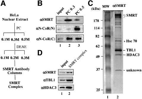
Fig. 2. Purification of a SMRT protein complex. (A) A diagram of the purification scheme for the SMRT complex. PC, phosphocellulose p11 resins; DEAE, DEAE–Sepharose Fast Flow resins. (B) Differential fractionation of SMRT and N-CoR proteins by a PC column. Note that αSMRT and αN-CoR(N) antibodies are specific for SMRT and N-CoR, respectively, whereas αN-CoR(C) antibody recognizes both SMRT and N-CoR. (C) A Coomassie blue-stained SDS–polyacrylamide gel showing the purified SMRT complex. The identity of the indicated subunits was determined by mass spectrometry and confirmed by western analyses as shown in (D). The Hsc70 protein was a contaminant because it also bound to the control IgG beads; 10% of the input (0.2 M DEAE fraction) was used in lane 1 in (D).

An official website of the United States government
Here's how you know
Official websites use .gov
A
.gov website belongs to an official
government organization in the United States.
Secure .gov websites use HTTPS
A lock (
) or https:// means you've safely
connected to the .gov website. Share sensitive
information only on official, secure websites.
