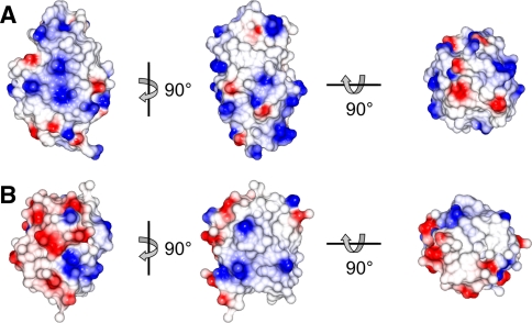
Fig. 3.
Surface representations of a HEWL (pdb code 193L (Vaney et al. 1996)) and b ubiquitin (pdb code 1UBQ (Vijay-Kumar et al. 1987), excluding the three flexible C-terminal residues) illustrating the overall shape of the proteins and their surface electrostatic potential (positive in blue, negative in red)

