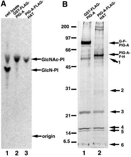
Fig. 1. Purification of GPI-GnT complexes. GST–FLAG-PIG-A and FLAG-HAT-PIG-A were isolated by two-step affinity purification from the digitonin lysates of 1.4 × 10⁸ cells of JY5 transfectants. Samples of the purified proteins (equivalent to 6 × 10⁷ cells) were used for in vitro GPI-GnT assay (A) and the rest (equivalent to 8 × 10⁷ cells) were used for SDS–PAGE and silver staining (B). (A) Lysates derived from 107 cells of wild-type JY25 as a positive control (lane 1) and purified complexes containing GST–FLAG-PIG-A (lane 2) or FLAG-HAT-PIG-A (lane 3) were incubated with radiolabeled UDP-GlcNAc and bovine PI. Lipids were analyzed by TLC. Identities of spots are indicated on the right. (B) Silver-stained SDS–PAGE profiles of purified complexes containing GST–FLAG-PIG-A (lane 1) and FLAG-HAT-PIG-A (lane 2). Band numbers are shown on the right. The positions of the molecular size markers are indicated on the left (kDa).

An official website of the United States government
Here's how you know
Official websites use .gov
A
.gov website belongs to an official
government organization in the United States.
Secure .gov websites use HTTPS
A lock (
) or https:// means you've safely
connected to the .gov website. Share sensitive
information only on official, secure websites.
