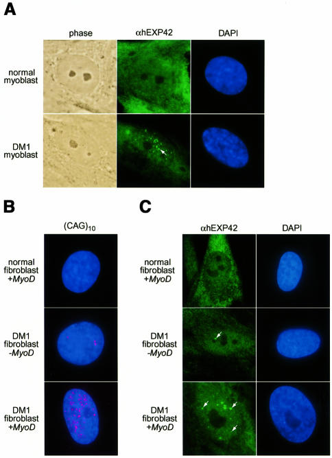
Fig. 6. EXP proteins accumulate in the nucleus of DM1 cells. (A) Cell immunofluorescence of normal and DM1 myoblasts using anti-EXP polyclonal antibodies. Phase contrast microscopy highlights cell position while DNA was detected with DAPI. The arrow indicates one of the EXP-enriched foci. (B) FISH analysis with a Cy3-labeled (CAG)10 probe (red), which detects DMPK (CUG)n expansions in DM1 cell nuclei (shown by DAPI staining). (C) Cell immunofluorescence, using anti-EXP antibodies, of normal fibroblasts infected with a MyoD adenovirus (normal fibroblast + MyoD) or DM1 fibroblasts either uninfected (DM1 fibroblast – MyoD) or infected (DM1 fibroblast + MyoD). Arrows indicate several sizes of foci enriched in hEXP42.

An official website of the United States government
Here's how you know
Official websites use .gov
A
.gov website belongs to an official
government organization in the United States.
Secure .gov websites use HTTPS
A lock (
) or https:// means you've safely
connected to the .gov website. Share sensitive
information only on official, secure websites.
