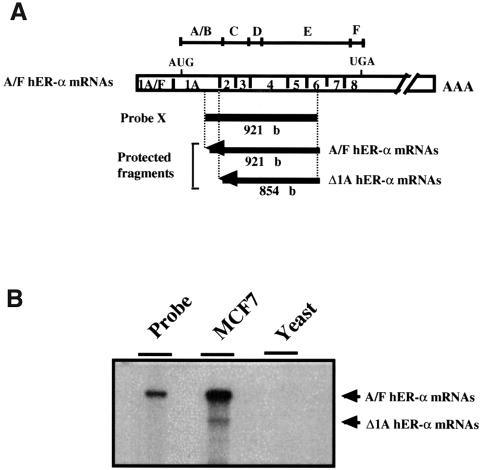
Fig. 1. Evidence for an alternative splicing event at exon 2 acceptor splice site of the hER-α gene. (A) Experimental design for Δ1A hER-α mRNA detection, indicating the location and the size of the single-stranded probe X and each protected fragment obtained after S1 digestion of probe/hER-α mRNA hybrids. Probe X (from +617 to +1538) was specific for normal hER-α transcripts (A/F hER-α mRNAs) but was also able partially to protect Δ1A hER-α mRNA isoforms up to the splice acceptor site position of exon 2. Open boxes indicate the unique (1A–F) and common (1–8) exons encoding each normal hER-α mRNA isoform. The positions of the initiator methionine (AUG) and the termination codon (UGA) are indicated. The division of the hER-α protein into six regions, A–F, is shown directly above the cDNA. (B) Total RNA (30 µg) from MCF7 cells and 30 µg of yeast RNA used as a negative control were hybridized to the labeled S1 probe X, treated with S1 nuclease, and the resistant hybrids were separated on a sequencing gel as described in Materials and methods. The undigested probe is shown in a separate lane.

An official website of the United States government
Here's how you know
Official websites use .gov
A
.gov website belongs to an official
government organization in the United States.
Secure .gov websites use HTTPS
A lock (
) or https:// means you've safely
connected to the .gov website. Share sensitive
information only on official, secure websites.
