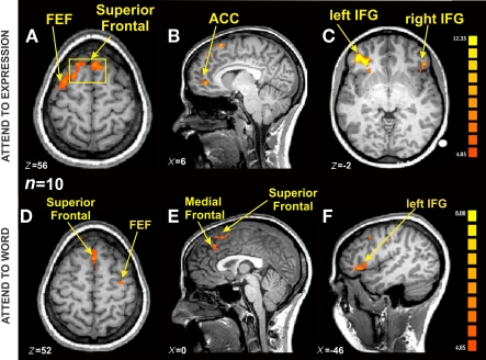
Figure 5.
fMRI BOLD activation from Incongruent blocks compared with Congruent blocks in the Expression and Word instruction conditions [n = 10, p(Bonferroni) < 0.05]. (A) Axial view showing the same Superior Frontal activation from (B) (yellow square), and a lateralized FEF activation on the left hemisphere (TAL coordinates: x = 5, y >= 22, z = 56). (B) Sagittal view centered on ACC on the right hemisphere (TAL coordinates: x = 6, y = 45, z = 1). (C) Axial view showing bilateral IFG activation (TAL coordinates: x = −39, y = 39, z = −2). (D) Axial view showing Superior Frontal activation, and a lateralized FEF activation on the right hemisphere (x = 0, y = −20, z = 52). (E) Sagittal view centered on Superior and Medial Frontal regions (TAL coordinates: x = 0, y = −9, z = 22). (F) Sagittal view centered on the left IFG (TAL coordinates: left IFG TAL coordinates: x = −46, y = 36, z = 4).

