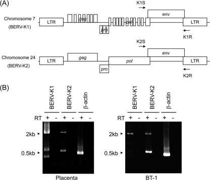
FIG. 1.
Genomic structures of BERV-K1 and -K2 identified by in silico analyses. (A) Schematic representation of BERV-K1 and -K2 proviruses on bovine chromosomes 7 and 24, respectively. Arrows indicate primers used for RT-PCR analysis in B. (B) Expression of BERV-K1 and -K2 mRNAs in bovine placenta and BT-1 cells. Total RNA was extracted from the placenta and BT-1 cells and then subjected to RT-PCR analysis using the primers indicated in A. β-Actin was also amplified as an internal control.

