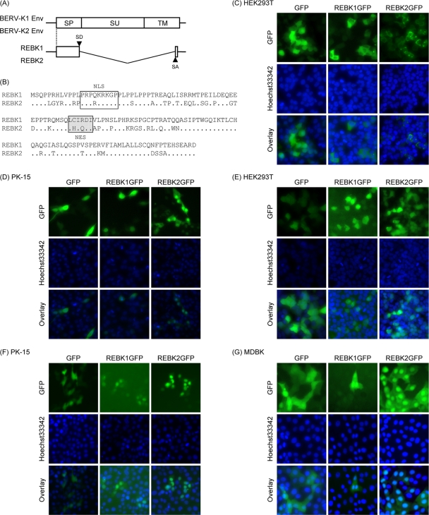
FIG. 4.
Identification of the REBK1 and REBK2 proteins. (A) Schematic representation of REBK1 and REBK2 transcripts. Start codons for REBK1 and REBK2 are shared with those of env. (B) Sequence alignment of the REBK1 and REBK2 proteins. Dots indicate identical amino acids. The box and the shaded box indicate NLS and NES motifs, respectively. (C to G) Subcellular localization of the REBK1 and REBK2 proteins. REBK1 and REBK2 fused with GFP (REBK1GFP and REBK2GFP, respectively) or only GFP (empty vector) was expressed in HEK293T (C and E), PK-15 (D and F), and MDBK (G) cells by transfection with expression plasmids (C and D) and infection with retroviral vectors (E, F, and G). Forty-eight hours after transfection and infection, the cells were stained with Hoechst 33342 dye and subjected to fluorescence microscopy analysis.

