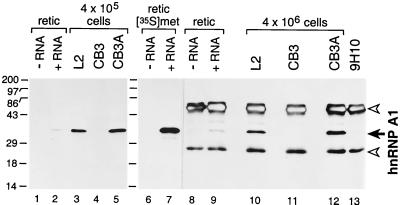
Figure 3.
CB3 cells produce no detectable hnRNP A1. hnRNP A1 (solid arrow) was detected in cell lysates by Western blotting (lanes 1–5) or by immunoprecipitation followed by Western blotting (lanes 8–13) with mAb 9H10. Blots were visualized by detection of chemiluminescence. Open arrowheads denote IgG heavy and light chains. As a control, rabbit reticulocyte lysate was programmed with synthetic RNA encoding hnRNP A1 (lanes 2, 7, and 9) or with H2O (lanes 1, 6, and 8) and labeled with [35S]methionine; lanes 6 and 7 are a fluorogram of lanes 8 and 9. Molecular masses (kDa) of marker proteins are indicated on the left.

