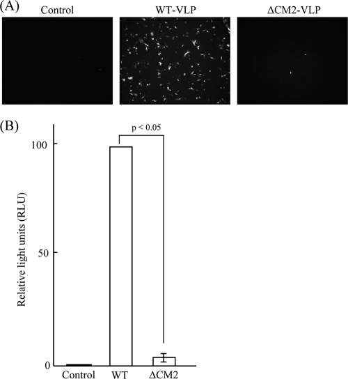
FIG. 3.
Reporter gene expression in HMV-II cells infected with VLPs. (A) HMV-II cells infected with mock (Control), WT VLPs, or ΔCM2 VLPs, followed by superinfection with AA/50, were incubated for 48 h and observed by fluorescence microscopy (magnification, ×100). (B) WT VLPs or ΔCM2 VLPs containing luciferase-vRNA were used for infection, and luciferase activity detected in the infected HMV-II cells was measured at 24 h p.i. and is expressed as relative light units (RLU). The RLU value from the HMV-II cells infected with WT VLPs was expressed as 100 and used for normalization. Each bar represents the mean ± standard errors of the means. The supernatant from mock-transfected 293T cells was used as a control.

