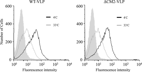
FIG. 4.
Flow cytometry of HMV-II cells infected with VLPs. HMV-II cells infected with WT VLPs or ΔCM2 VLPs were analyzed by flow cytometry using anti-HEF MAb J14. The VLP-infected cells were incubated at 4°C for 30 min and then incubated at 33°C for a further 180 min. The histogram from mock-infected cells is shown as a shaded area. Vertical and horizontal lines indicate the number of cells and fluorescence intensities, respectively.

