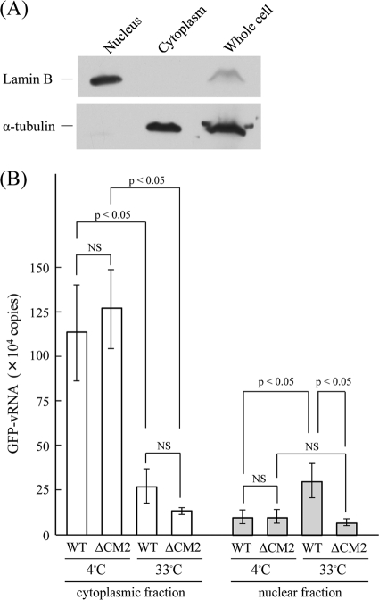
FIG. 6.
Fractionation and real-time PCR of HMV-II cells infected with VLPs. (A) HMV-II cells were fractionated as described in Materials and Methods, and the whole-cell lysates, the nuclear fraction, and the cytoplasmic fraction of the cells were analyzed by immunoblotting using anti-lamin B and anti-α-tubulin antibodies. (B) The HMV-II cells infected with WT VLPs or ΔCM2 VLPs were incubated at 4°C for 30 min and then transferred to 33°C and incubated for 60 min. The cells were divided into cytoplasmic and nuclear fractions, and the GFP-vRNA contained in the respective fractions was quantified by real-time PCR. The vertical line indicates the copy number of GFP-vRNA from 1.0 × 10⁶ HMV-II cells infected with VLPs. The data obtained from three independent experiments are shown as the means ± standard deviations. All comparisons between groups were statistically evaluated by using a paired t test. NS, not significant.

