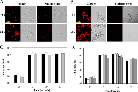
FIG. 3.
Copper uptake into cells exposed to copper surfaces. Cells of C. albicans (A, C) or S. cerevisiae (B, D) were exposed at 23°C to pure copper or to stainless steel (SS) for the indicated times, removed, washed, stained with the Cu(I)-specific fluorescent dye coppersensor-1, and subjected to fluorescence microscopy (A, B) or were mineralized and subjected to ICP-MS analysis for determination of cellular copper content (C, D). Bars represent wild-type C. albicans (black), a Cacrp1Δ/Cacrp1Δ mutant lacking a copper efflux P-type ATPase (gray), or a Cacup1Δ/Cacup1Δ mutant lacking a copper metallothionein (white) (C) and wild-type S. cerevisiae (black), an Scctr1Δ mutant lacking a copper uptake permease but harboring a control plasmid (white), an Scctr1Δ mutant complemented with ScCTR1 (light gray), an Scctr1Δ mutant complemented with hyperactive Scctr1(300) (dark gray), or an ace1Δ mutant not expressing metallothioneins (medium gray) (D). Shown are fluorescence (left) and representative phase contrast (right) microscopy images (A, B) or averages of results from triplicate experiments, with standard deviations (error bars) (C, D).

