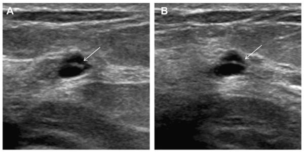
Fig. 10.
(A) Transverse and (B) sagittal ultrasonographic images (L12-5 MHz transducer) of a 47-year-old woman with incidental cyst containing a thin (<0.5 mm) septation (arrows) on ACRIN 6666 screening ultrasonography, which was stable for 3 years. This is a benign finding. (Courtesy of Wendie A. Berg, MD, PhD, Lutherville, MD.)

