
Fig. 14.
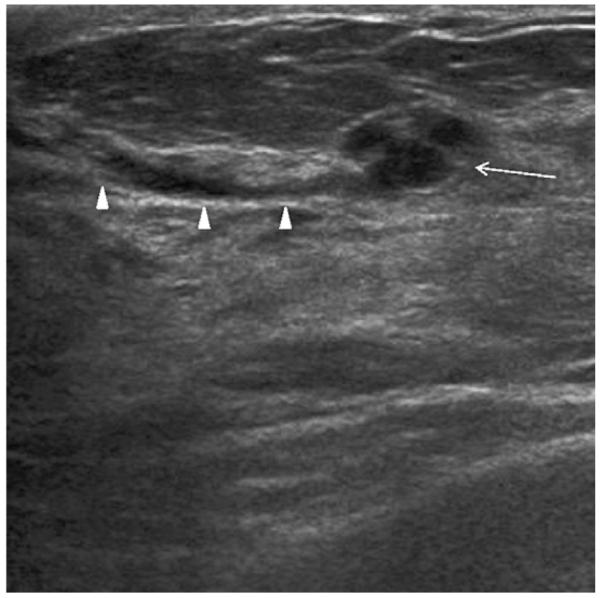
Radial ultrasonographic image shows incidental group of small cysts (arrow) communicating with a duct (arrowheads) in a 51-year-old woman who underwent screening ultrasonography due to a strong family history of breast cancer and dense breasts. (Courtesy of Wendie A. Berg, MD, PhD, Lutherville, MD.)