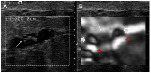
Fig. 17.
A 40-year-old woman was found to have a mass on screening mammography. (A) Targeted ultrasonography showed an intraductal mass (arrow), which was hypervascular on power Doppler (not shown). (B) Elasticity image shows bull’s-eye appearance to the fluid (solid arrows) on either side of the solid, gray, stiffer, biopsy-proven papilloma (white arrow). Posterior bright areas (red arrows) are also noted deep to the cystic portions. (Courtesy of Richard G. Barr, MD, PhD, Radiology Consultants, Youngstown, OH.)

