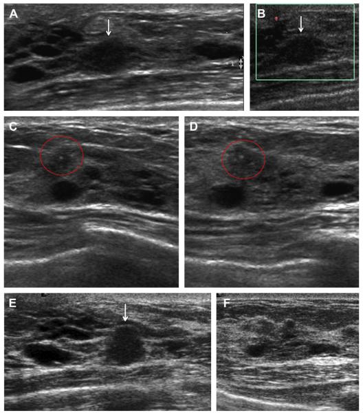
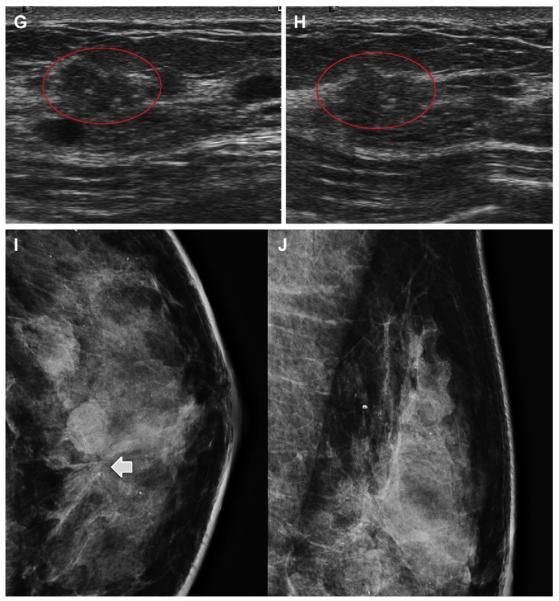
Fig. 23.
A 52-year-old ACRIN 6666 participant had multiple findings on her third annual screening ultrasonography, including multiple cysts. On (A) radial and (B) antiradial ultrasonography with power Doppler, a mass seen at 2-o’clock position in the left breast was thought to be a complicated cyst and was recommended for a 6-month follow-up. On (C) radial and (D) antiradial ultrasonographic images of the right breast, at the 12-o’clock position, a few calcifications were noted (red circles), which were dismissed as fibrocystic changes adjacent to several small cysts. No calcifications were seen mammographically. At follow-up after 7 months, (E) radial ultrasonography showed that the left breast mass had enlarged (arrow), prompting ultrasound-guided biopsy. This mass proved to be a 6-mm node-negative invasive lobular carcinoma. (F) Radial ultrasonography of the 12-o’clock position in the left breast appeared unremarkable. Follow-up (G) transverse and (H) sagittal ultrasonography on the right side showed an enlarging isoechoic mass with calcifications (red ovals). This mass proved to be a 6-mm grade I IDC+DCIS and node negative. Seen only on (I) CC and not on (J) MLO mammograms, a subtle area of distortion was noted in the 12-o’clock position of the left breast (short solid arrow), which proved to be a 5-mm tubular carcinoma (grade I IDC, special type) with associated DCIS. (Courtesy of Wendie A. Berg, MD, PhD, Lutherville, MD.)


