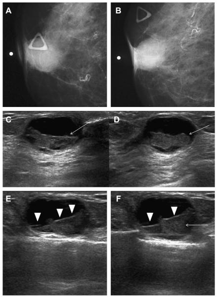
Fig. 27.
A 68-year-old woman was noted to have a palpable mass at the nipple, marked by triangular markers on (A) spot compression CC and (B) MLO mammograms that show the palpable abnormality to correspond to a dense indistinctly marginated mass with overlying skin retraction. (C) Radial and (D) antiradial ultrasonography show intracystic mass (arrows). Additional oblique ultrasonographic images (E, F) show fluid-debris level (arrowheads) formed due to hemorrhage from the intracystic mass (arrow). A 14-gauge core biopsy showed atypical papilloma. Excision showed 8 mm of DCIS involving a papilloma, with 3-mm microinvasive colloid carcinoma. (Courtesy of Wendie A. Berg, MD, PhD, Lutherville, MD.)

