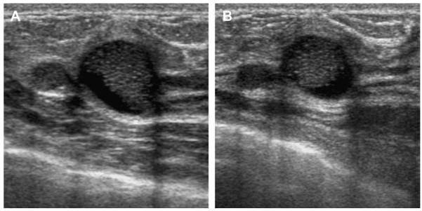
Fig. 29.
On screening ACRIN 6666 ultrasonography, a 54-year-old woman was noted to have a circumscribed oval mass with posterior enhancement in the outer right breast on (A) supine position. Internal echoes were noted, and it was uncertain whether the echoes represented debris or an intracystic mass. (B) The patient was then positioned in left lateral decubitus (LLD) position and reimaged after 3 minutes, showing a shift in the contents, which is consistent with debris. This is a benign complicated cyst with no evidence of an intracystic mass. (Courtesy of Wendie A. Berg, MD, PhD, Lutherville, MD.)

