Fig. 41.
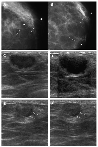
A 37-year-old woman is 18 months postreduction mammoplasty and noted 2 palpable lumps (marked with radiopaque markers). On close-up (A) CC and (B) MLO mammograms, the masses are shown to be circumscribed and lucent, typical of benign oil cysts (arrows). Ultrasonography was nonetheless performed because of other abnormalities. (C) Transverse and sagittal ultrasonographic images of the mass in the 6-o’clock position show a nearly anechoic mass with thick nodular wall and no posterior features. Without the mammogram, the sonographic appearance would be indeterminate. (E) Radial and (F) antiradial ultrasonographic images of the oil cyst at 12-o’clock position show it to be mostly (but not completely) circumscribed, nearly isoechoic to surrounding fat, and to contain a thin eccentric rim of fluid (arrows). (Courtesy of Wendie A. Berg, MD, PhD, Lutherville, MD.)

