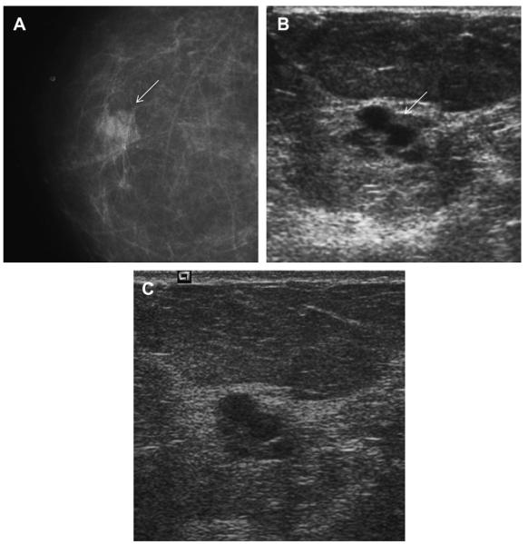
Fig. 52.
A 78-year-old woman was noted to have a new mass on screening mammography, as seen on (A)CC mammogram (arrow). (B) Targeted ultrasonography demonstrated what appeared to be clustered microcysts (arrow). The patient was not on hormonal therapy. Short-interval follow-up was recommended. At (C) 6-month follow-up ultrasonography, the mass seemed to have thick septations and indistinct margins. Biopsy was recommended, showing grade III IDC with associated high nuclear grade DCIS. (Courtesy of Wendie A. Berg, MD, PhD, Lutherville, MD.)

