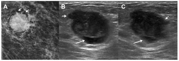
Fig. 54.
A 74-year-old woman noted a lump in her left breast. (A) Spot compression mammogram demonstrated a mostly circumscribed dense mass. A portion of the margin appears lobulated (short arrows). Targeted (B) radial and (C) antiradial ultrasonography demonstrated a complex mostly solid mass with eccentric cystic area (arrows). The margins are focally angular (short arrows). Ultrasound-guided biopsy showed grade III IDC. (Courtesy of Wendie A. Berg, MD, PhD, Lutherville, MD.)

