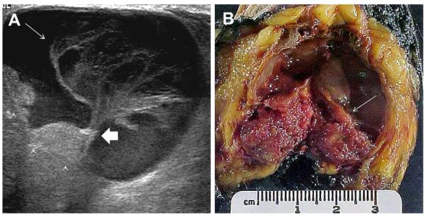
Fig. 64.
A 75-year-old woman noted a palpable mass. On (A) targeted ultrasonography, a complex cystic and solid mass was noted with a frondlike appearance to the intracystic mass (long arrow). A vascular stalk (short fat arrow) was noted to the intracystic mass, and the wall was focally thickened (arrowheads). (B) Gross specimen demonstrates the intracystic mass (arrow). Histopathology showed in situ papillary carcinoma, which was focally invasive where the wall was thickened. (Courtesy of Dr Eva Gombos, Brigham and Women’s Hospital, Boston, MA.)

