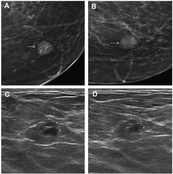
Fig. 66.
A 45-year-old woman with prior history of fibroadenoma at 12-o’clock position in the left breast had an enlarging circumscribed mass in the lower inner left breast on screening (A) CC and (B) MLO (arrows). Targeted (C) radial and (D) antiradial ultrasonography demonstrated an isoechoic oval mass (arrows) with small eccentric cystic areas (arrowheads), that is, a complex cystic and solid mass. Ultrasound-guided 14-gauge core biopsy confirmed a complex fibroadenoma with usual duct hyperplasia and cystic areas. (Courtesy of Wendie A. Berg, MD, PhD, Lutherville, MD.)

