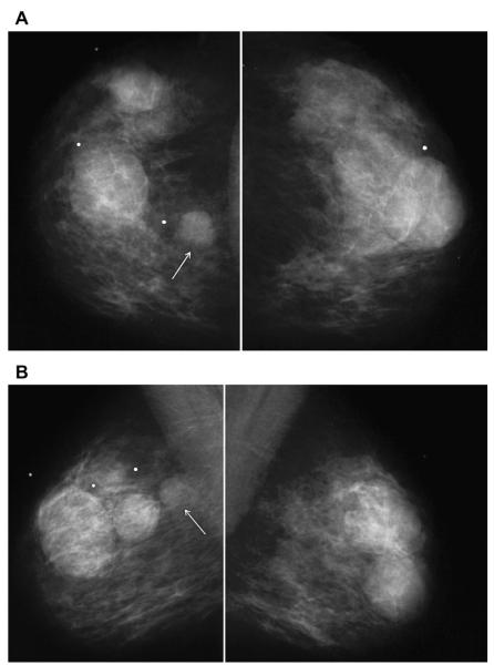
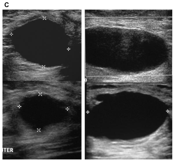
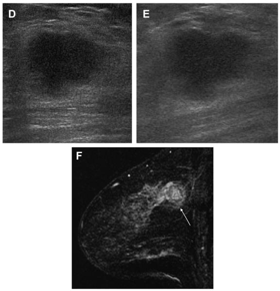
Fig. 7.
A 45-year-old woman had a history of cysts and now had bilateral tender palpable masses marked by radiopaque markers on (A) CC and (B) MLO mammograms. (C) Multiple static ultrasonographic images of the palpable lumps were interpreted as bilateral cysts. One lump was aspirated (upper inner right breast [marked by arrows in A and B]), yielding malignant cytology. (D) Repeat ultrasonography of the known malignant mass in the right breast at 2-o’clock position shows it to be indistinctly marginated and lobulated both without and (E) with spatial compounding. Any lesion documented on ultrasonography should have images recorded without calipers to facilitate margin assessment. Images of the tumor from the initial ultrasonography in part C (the bottom left image) had too narrow a dynamic range (effectively too few shades of gray); hence, the solid mass appeared anechoic. (F) Sagittal image from 3-dimensional fat-suppressed spoiled gradient echo (SPGR) MR imaging 1.5 minutes after 0.1 mmol/kg of gadolinium-based contrast injection shows an irregular enhancing mass (arrow) corresponding to the known grade III invasive ductal carcinoma. (Courtesy of Wendie A. Berg, MD, PhD, Lutherville, MD.)


