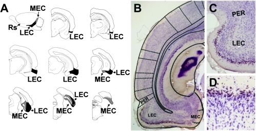
Figure 2.
Reelin is a marker expressed by neurons in the superficial layers of the lateral entorhinal cortex. (A) Sagittal view of the rat brain, showing the anterior and inferior anatomical position of the lateral entorhinal cortex (LEC) and the posterior situation of the medial entorhinal cortex (MEC). The LEC is visible on coronal sections containing the dorsal hippocampus. As sections become more caudal, the LEC occupies more of the temporal cortex. On caudal sections that do not contain hippocampus, the MEC is visible at the medial edge of the cortex. Eventually, the MEC ascends to encompass the temporal cortical field. Image in (A) adapted from Rapp et al. (2002). (B) Low-magnification image showing reelin immunoreactivity in the superficial layers of the LEC. The diagram superimposed on the image is from Paxinos and Watson (1998). (C) Reelin staining in layer II is confined to the LEC, located inferior to the rhinal sulcus on coronal sections. No reelin staining is detected in layer II of perirhinal cortex (PER) surrounding the superficial portion of the rhinal sulcus. (D) Immunoreactivity for reelin among neurons in layer II of the lateral entorhinal cortex.

