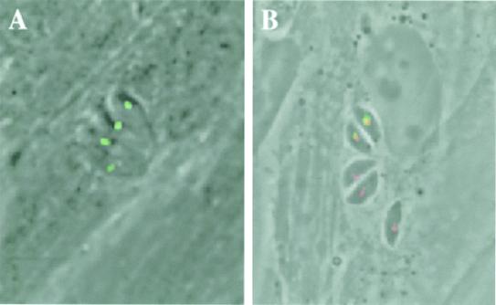
Figure 3.
T. gondii ACC1 is localized in the apicoplast. Fusing GFP to the putative N-terminal-targeting signal of ACC1 was sufficient to direct this reporter to the apicoplast in living parasites (A). Subcellular localization of GFP was assessed by direct fluorescence microscopy of live parasites 24 h after transient transfection. The apicoplast ACC1 localization was confirmed in fixed cells by localization of ACP with anti-ACP antibodies and GFP with anti-GFP antibodies, followed by detection with rhodamine- and FITC-conjugated secondary antibodies, respectively (B). Three parasitophorous vacuoles are visible in B: the upper vacuole (containing two parasites) was transiently transfected with ACC1-GFP; the yellow signal indicates precise colocalization with ACP.

