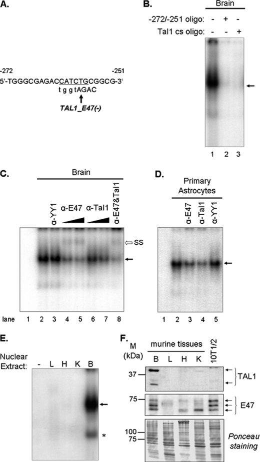
FIGURE 4.
Binding of Tal1 and E47 to the mouse promoter. A, nucleotide sequence of the −272/−251-bp fragment showing the putative E47/Tal1 element. The minus strand is indicated by a minus sign. B–D, competition and supershift assays of proteins binding to the promoter were performed using 32P-labeled −272/−251-bp sequence as probe. B, nuclear extracts from brains of 1-day-old mice incubated with: probe alone (lane 1), unlabeled −272/−251-bp fragment (lane 2), or Tal1 consensus (cs) binding sequence (lane 3). Oligonucleotides were added at 100-fold molar excess relative to probe for 10 min before incubation with probe. C, probe alone (lane 1); nuclear extract from brains of 1-day-old mice was incubated with probe (lane 2) or preincubated with anti-YY1 antibody (lane 3), anti-E47 antibody (lanes 4 and 5), anti-Tal1 antibody (lanes 6 and 7), or both antibodies (lane 8), before addition of probe. D, probe alone (lane 1), nuclear extract from mouse primary astrocytes was incubated with probe (lane 2) or preincubated with anti-E47 (lane 3), anti-Tal1 (lane 4), or anti-YY1 (lane 5) antibodies. E, nuclear extracts (20 μg of protein) from brain (B), liver (L), heart (H), and kidney (K) of 1-day-old mice were used. Arrow, E47-Tal1/DNA complex; *, complex (presumably truncated Tal1 with E47) visible only when at least 20 μg of protein were used. F, nuclear extracts from brain (B), liver (L), heart (H), and kidney (K) of newborn mice were immunoblotted with antibodies against E47 and Tal1. Nuclear extract from C3H10T1/2 cells (10T1/2) was used as negative control. Ponceau Red staining indicates similar loading of proteins in each lane. M, molecular mass (kDa). Each experiment was repeated three to five times with similar results.

