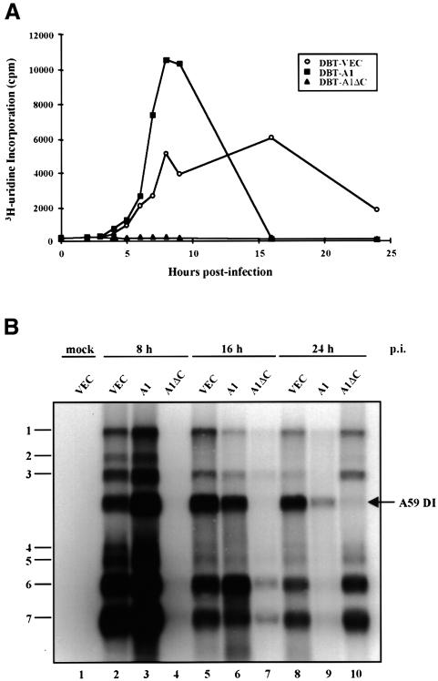
Fig. 5. Kinetics of MHV RNA synthesis in DBT cells. (A) [3H]uridine labeling of MHV RNA in DBT cells. DBT-VEC, DBT-A1 and DBT-A1ΔC cells were infected with MHV-A59 at an m.o.i. of 2. At 1 h p.i., serum-free medium was replaced by virus growth medium containing 1% NCS and 5 µg/ml actinomycin D. [3H]uridine (100 µCi/ml) was added to the infected cells at 2, 3, 4, 5, 6, 7, 8, 9, 16 and 24 h p.i. After 1 h labeling, cytoplasmic extracts were prepared and precipitated with 5% TCA. The TCA-precipitable counts were measured in a scintillation counter. (B) Northern blot analysis of MHV genomic and subgenomic RNA synthesis in DBT cells. Cytoplasmic RNA was extracted from MHV-A59-infected cells at 8, 16 and 24 h p.i. for northern blot analysis. The naturally occurring DI RNA of MHV-A59 is indicated by an arrow.

An official website of the United States government
Here's how you know
Official websites use .gov
A
.gov website belongs to an official
government organization in the United States.
Secure .gov websites use HTTPS
A lock (
) or https:// means you've safely
connected to the .gov website. Share sensitive
information only on official, secure websites.
