FIGURE 2.
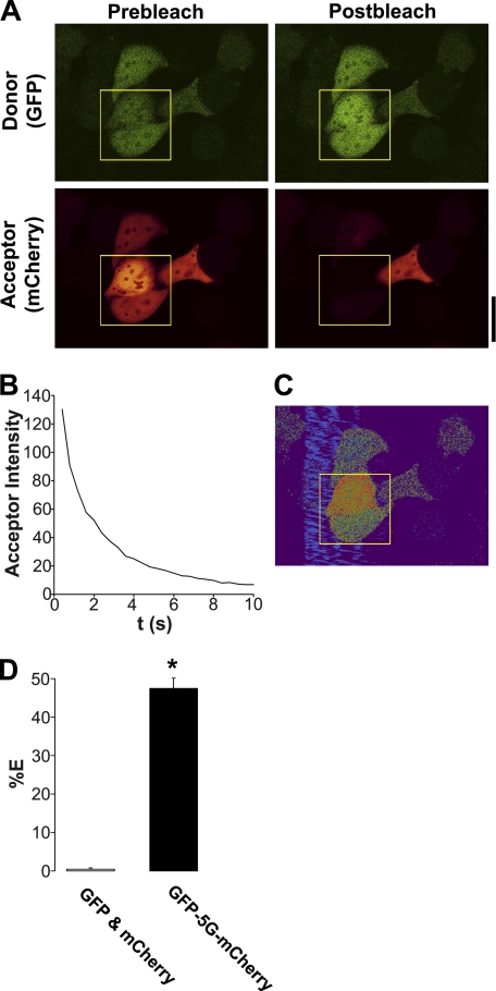
eGFP linked to mCherry with 5 glycines (eGFP-5G-mCherry) undergoes near-maximal FRET. A, typical confocal micrographs of eGFP-5G-mCherry (green, donor; red, acceptor) before and after photobleaching of the acceptor fluorophore. B, graph showing typical acceptor photobleaching over 10 s. C, resultant FRET image from A. D, mean FRET efficiency (%E) of unlinked eGFP and mCherry cotransfected in the same cells (n = 10) and from eGFP-5G-mCherry (n = 14). * denotes p < 0.05 different between linked and unlinked eGFP and mCherry. Scale bar represents 25 μm.

