FIGURE 1.
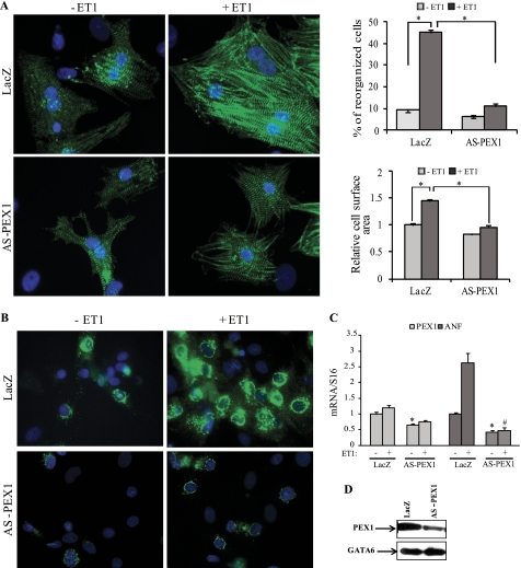
PEX1 is required for ET-1 signaling. A, left, Hoechst (blue) and sarcomeric α-actinin (green) immunofluorescence-labeled slides are shown of the control adeno-LacZ and adeno-HA-AS-PEX1-infected primary cardiomyocytes treated or not with 100 nm ET-1 for 24 h. Costaining was performed using Hoechst to detect cell nuclei and sarcomeric α-actinin to visualize the myofibrils of cardiomyoctes. Note that the loss of PEX1 blocked the myofibrillar reorganization seen in adeno-LacZ-infected cells treated with ET-1. A, right, reorganized cells were counted, and relative cell surface area was measured across 10 fields (×40) in three separate experiments. *, p < 0.05. B, ANF immunofluorescence is shown in cardiomyocytes infected for 3 days with either adeno-LacZ or adeno-HA-AS-PEX1 and treated with vehicle or ET-1. Note how PEX1 down-regulation inhibited ET-1-induced cellular accumulation of ANF. C, ANF mRNA levels in LacZ- or AS-PEX1-transfected cardiomyocytes treated or not with ET-1. *, p < 0.05 versus LacZ; #, p < 0.05 versus LacZ + ET-1. D, PEX1 protein levels in cardiomyocytes infected with the adeno-AS-PEX1 or adeno-LacZ as detected by Western blotting are shown. GATA6 protein was used as an internal control.

