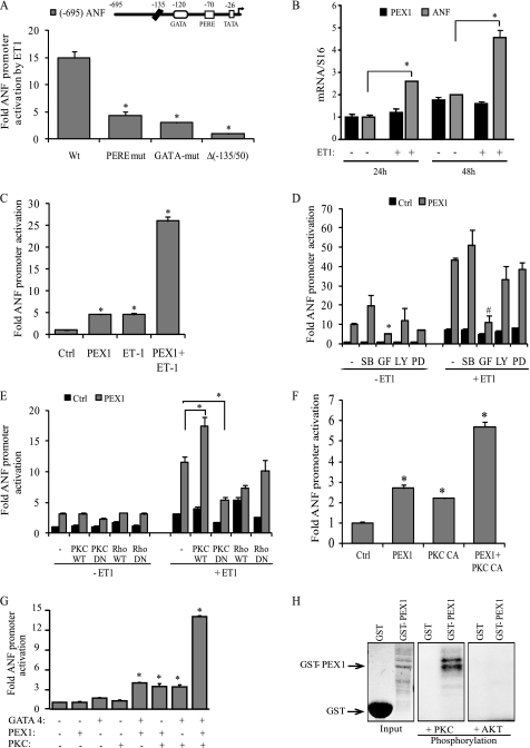
FIGURE 2.
Post-translational regulation of PEX1 by ET-1. A, ANF promoter elements required for ET-1 response in cardiomyocytes. Cells were transfected with wild-type (Wt) and mutated −695ANF or −135ANF promoter luciferase reporter constructs and stimulated for 48 h with 100 nm ET-1. PERE mut corresponds to mutation of the proximal PERE site (position −70 bp); GATA mut corresponds to mutation of the GATA site (position −120 bp). The data are mean of ± S.E. (error bars; n = 4). *, p < 0.05 versus wild type. B, effect of ET-1 on PEX1 mRNA levels. ANF and PEX1 transcript levels were quantified in primary cultured ventricular cardiomyocytes treated or not with 100 nm ET-1 for 24 or 48 h. Data are mean ± S.E. (n = 4). *, p < 0.05 C, ET-1 potentiates PEX1 activation. Cardiomyocytes were cotransfected with the proximal ANF promoter luciferase reporter and 500 ng of PEX1. The cells were then treated with 100 nm ET-1 for 48 h. Data are mean ± S.E. (n = 4). *, p < 0.05 versus Ctrl. D, involvement of PKC but not p38-MAPK or ERK1/2 in ET-1-dependent PEX1 activation of ANF transcription. Cardiomyocytes were cotransfected with the proximal ANF promoter luciferase reporter and 500 ng of PEX1. The cells were then treated with 100 nm ET-1 for 24 h in the presence of inhibitors: p38 MAPK (SB 203580; 10 μm [SB]), PKC (GF 109203X; 5 μm [GF]), PI3K (LY 294002; 25 μm [LY]) ERK1/2 (PD98059; 10 μm [PD]). The data are from one representative experiment carried out in triplicate. *, p < 0.05 versus PEX1 (−ET1); #, p < 0.05 versus PEX1 (+ET1). E and F, cotransfection in cardiomyocytes of the ANF-Luc reporter with the indicated expression vectors treated or not with 100 nm ET-1(PKC CA, PKCβ catalytic domain). Note how cotransfection with PKC-dominant negative (DN) but not Rho-DN abrogates the ET-1 effect on PEX1 activation of the promoter. *, p < 0.05. G, PKCβ synergizes with PEX1 and GATA4 on the ANF promoter. NIH3T3 cells were cotransfected with the ANF-Luc construct 100 ng of PEX1, 10 ng of GATA4, and 200 ng of PKCβ CA. *, p < 0.05 versus Ctrl. H, in vitro PKC phosphorylation of GST-PEX1 fusion protein. In similar kinase assays (such as AKT kinase), PEX1 was not phosphorylated, demonstrating PKC specificity. Coomassie staining was used to show protein loading.

