FIGURE 4.
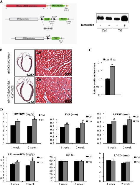
Conditional overexpression of PEX1 in adult mice hearts leads to cardiac hypertrophy. A, strategy used for conditional expression of PEX1 using the Cre/LoxP system. α-MHCMerCreMer mice were crossed with CAT-PEX1 transgenic mice to obtain a conditional transgenic line that overexpresses PEX1 strictly in cardiomyocytes once Tamoxifen is administered. Right, transgenic mice receiving tamoxifen showing increased PEX1 protein levels, whereas those injected with peanut oil and controls receiving tamoxifen showing no change. B, image magnified ×1.25 of trichrome-stained heart sections from 150-day-old α-MHCMerCreMer or α-MHCMerCreMer/CAT-PEX1 mice treated with Tamoxifen. Notice the hypertrophied heart when PEX1 expression is induced. Magnification ×63 shows increased myocyte size. C, relative cell surface area was measured across 10 fields (×40) in transgenic (TG) and control (Ctrl) mice (2 of each). *, p < 0.05 versus control. D, echocardiographic analysis of mice hearts 1 week and 2 weeks after Tamoxifen administration. Heart weight-to-body weight ratios (HW/BW) are also shown. IVS, interventricular septum thickness; LVPW, left ventricular posterior wall thickness; LVID, left ventricular internal dimension; LV mass, left ventricular mass; EF, ejection fraction. Notice how ejection fraction was not changed despite the increase in left ventricular mass/body weight ratio suggestive of preserved heart function. The data are means ± S.E. (error bars; n = 4–5 for each group). *, p < 0.05 versus control (Ctrl; 1 week); #, p < 0.05 versus control (2 weeks).

