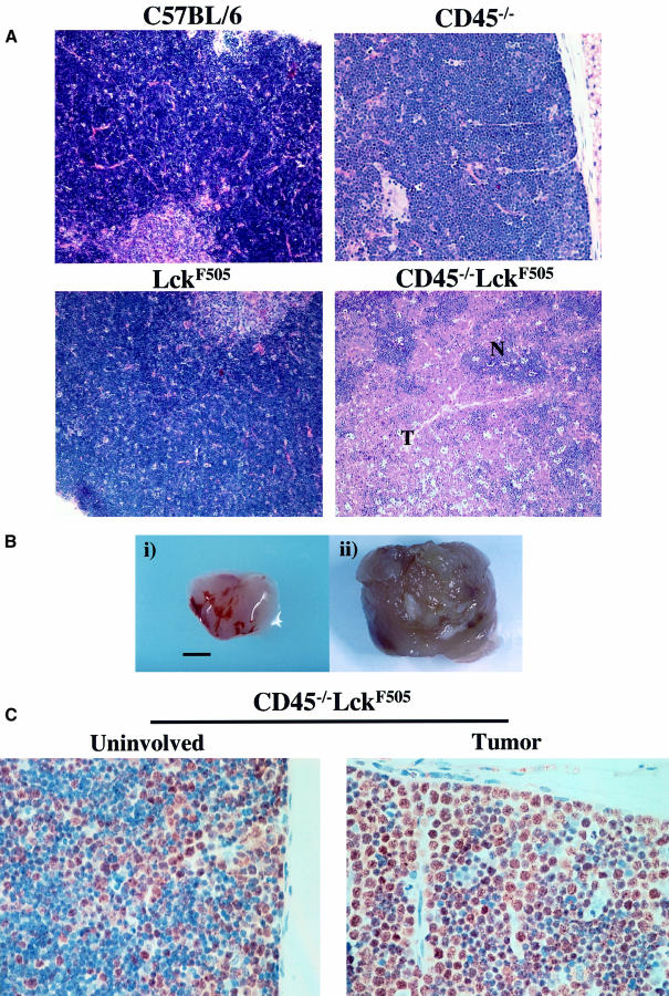
Fig. 2. CD45–/–LckF505 mice develop thymic lymphoblastic lymphomas. (A) Hematoxylin and eosin-stained thymic sections from a 7-week-old mouse displaying representative early tumour development are shown with CD45–/–, LckF505 and C57BL/6 thymus for comparison. Early transformation events characteristically show lymphoblastic cells developing in foci (T) surrounded by normal tissue (N) often localized to one lobe of the thymus. (B) Comparison of normal (i) C57BL/6 and transformed (ii) CD45–/–LckF505 thymuses. The scale bar represents 5 mm. (C) A thymic tumour from a 7-week-old mouse in which proliferating (brown stain) lymphoblastic cells are amplified in one lobe (‘tumor’), the other lobe (‘uninvolved’) displaying a wild-type morphology.

An official website of the United States government
Here's how you know
Official websites use .gov
A
.gov website belongs to an official
government organization in the United States.
Secure .gov websites use HTTPS
A lock (
) or https:// means you've safely
connected to the .gov website. Share sensitive
information only on official, secure websites.
