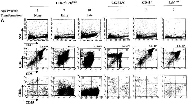
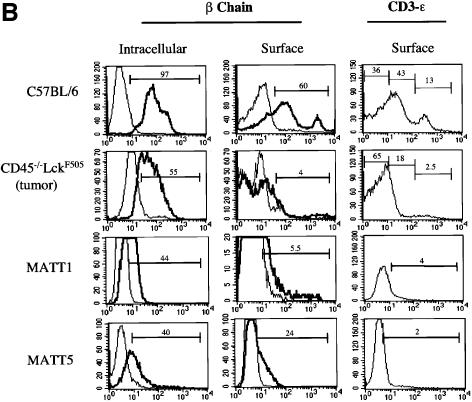
Fig. 3. Thymic lymphomas in CD45–/–LckF505 mice originate from immature thymocytes expressing abnormal levels of TCR-β and CD3-ε. (A) Thymic expression of CD4, CD8, CD25 and CD44 cell surface antigens was analysed by flow cytometry in 7-week-old CD45–/–LckF505 litter-mate mice that did (‘early’) or did not (‘none’) display developmental defects, and in a 10-week-old mouse bearing a fully developed lymphoma. Wild-type C57BL/6, CD45–/– and LckF505 (PLGF-A) thymocytes are shown for comparison. Values above the CD4 versus CD8 plots represent total cell numbers from each thymus, whereas values in each quadrant represent the percentage of cells in that quadrant. (B) TCR-β and CD3-ε expression in C57BL/6 thymocytes, primary CD45–/–LckF505 lymphoma cells and in two cell lines (MATT-1 and MATT-5) established from tumours derived from two different CD45–/–LckF505 mice. TCR-β chain plots show cells stained using TCR-β–FITC (bold line) or an irrelevant isotype control mAb (thin line). CD3-ε staining was carried out using CD3-ε–FITC. The values on the plots represent percentage positive cells within the gate indicated.

An official website of the United States government
Here's how you know
Official websites use .gov
A
.gov website belongs to an official
government organization in the United States.
Secure .gov websites use HTTPS
A lock (
) or https:// means you've safely
connected to the .gov website. Share sensitive
information only on official, secure websites.

