FIGURE 4.
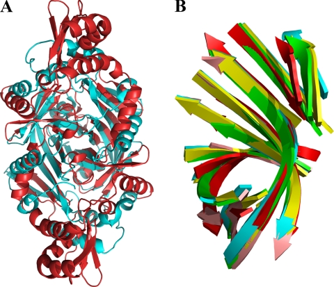
Structural similarities between HugZ and FMN-binding split-barrel proteins. A, superposition of HugZ and a randomly selected FMN-binding split-barrel protein (Protein Data Bank code 1TY9). HugZ is colored red, and the FMN-binding protein is cyan. Heme and FMN are removed for clarity. B, superposition of β-strand substructures of the HugZ monomer (red, C-terminal domain only) with four randomly chosen FMN-binding split-barrel proteins (1CI0, 1DNL, 1NRG, and 1RFE).

