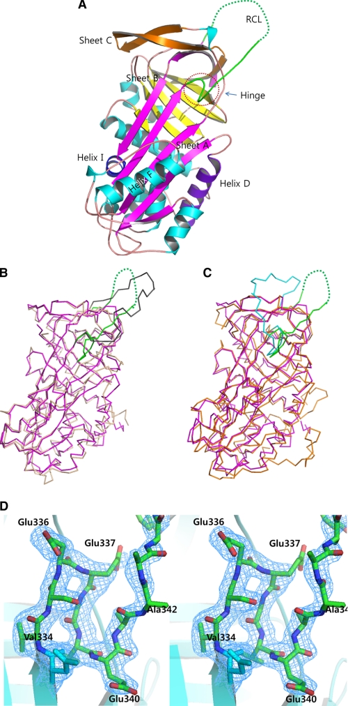
FIGURE 2.
Overall structure of SPN48. A, ribbon representation of the SPN48 structure. The RCL is shown in green. Helices are in cyan; helix D is in purple, and helix I is in blue. Sheet A is shown in magenta, and sheets B and C are in yellow and orange, respectively. B, superposition of SPN48 (green RCL and magenta elsewhere) and serpin 1K (gray RCL and lime elsewhere) using Cα atoms. Structural superposition was carried out using PyMOL (29). C, superposition of SPN48 (green RCL and magenta elsewhere) and antithrombin (cyan RCL and orange elsewhere) using Cα atoms. Structural superposition was carried out using PyMOL (29). D, stereo representation of the hinge region of SPN48 with the electron density map. 2FoFc electron density map is contoured at 1.0σ level.

