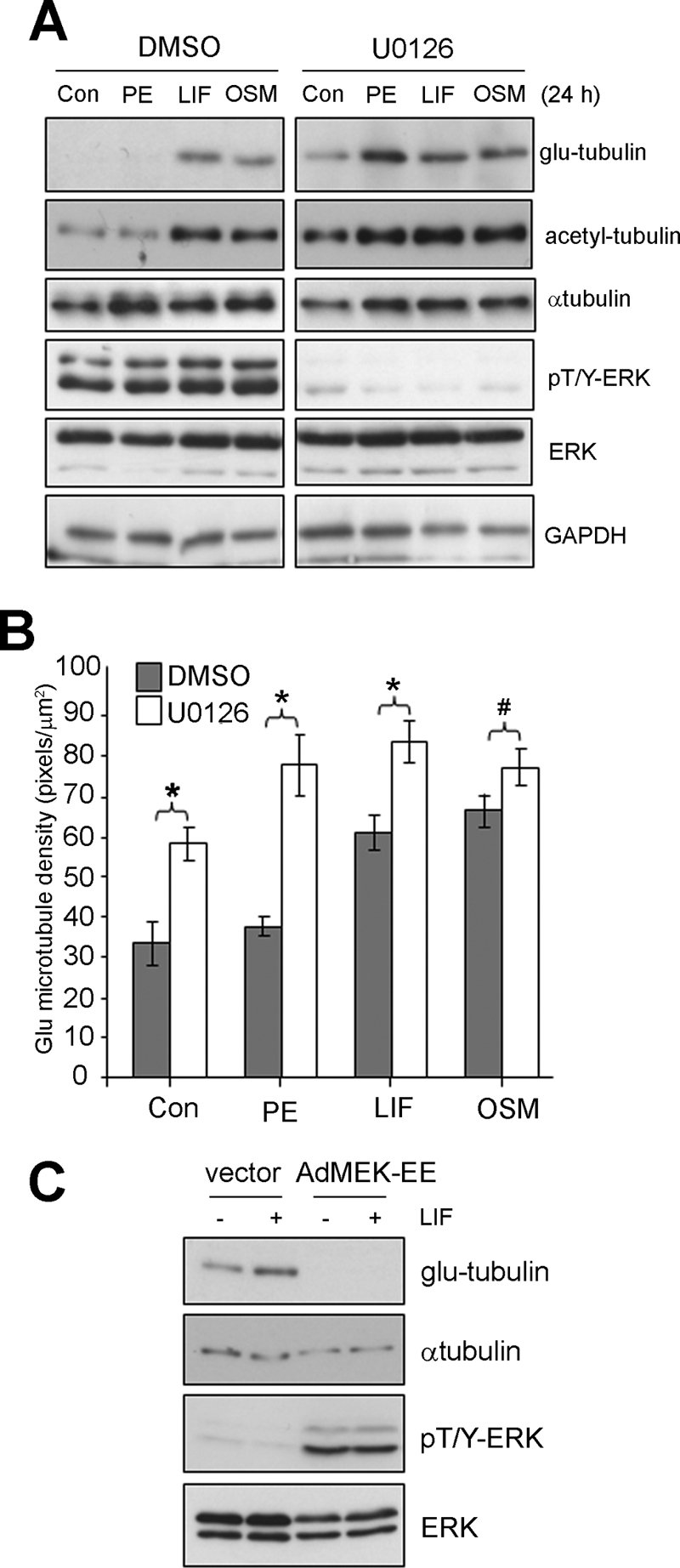
FIGURE 4.

MEK/ERK signaling negatively regulates stable MTs in cardiac myocytes. Cultured cardiac myocytes were pretreated with U0126 (10 μm, 30 min) or a vehicle control (0.1% (v/v) DMSO), then stimulated (24 h) with agonists, PE (100 μm), LIF (10 ng/ml), OSM (10 ng/ml), or 0.1% (w/v) BSA in PBS as a control. A, Protein lysates were then prepared blotted with the indicated antibodies. B, Glu-MTs were immunostained to revealed the stabilized MT network and the density of glu-MTs quantitated by immunofluorescence image analysis and results expressed as an average ± S.E. (*, p < 0.01, #, p < 0.05, n = 3). C, cardiac myocytes were infected (50 moi) with adenovirus encoding MEK-EE (AdMEK-EE) or a shuttle vector only control (vector) before stimulating with LIF (10 ng/ml, 24 h) or equivalent volume of 0.1% BSA (w/v) in PBS. Protein lysates were then prepared and blotted with the indicated antibodies.
