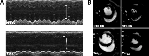
FIGURE 7.
Impaired cardiac function in 8-month-old taz-deficient (Tazdef) mice. A, representative M-mode echocardiographic tracings of NTG and taz-deficient mice. Diastolic dimension is indicated by solid lines, and systolic dimension is indicated by dotted lines. Chamber dilation and thinning of the ventricular walls are evident in the taz-deficient mouse. B, representative short axis cardiac MRI of NTG and taz-deficient mice at end diastole (ED) and end systole (ES).

