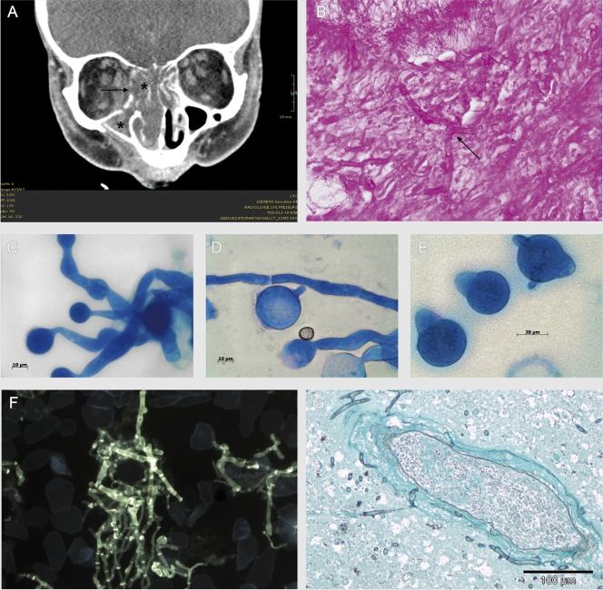
FIG. 1.
(A) CT scan of the midface: signs of sinusitis (*) and osteolyses of the medial part of the right orbita (→). (Courtesy of Mathias Langer and Marisa Windfuhr-Blum, Department of Radiology, University Hospital of Freiburg; reproduced with permission.) (B) Hyphae with orthogonal branches in periodic acid-Schiff staining (magnification, ×600) in the biopsy specimens of the ethmoidal cells. (C to E) Micromorphology of Conidiobolus incongruus (lactophenol blue; magnification, ×1,000). (C and D) Wide vegetative mycelium with moderate septation. (D and E) Large single-celled primary conidia with pointed papillae. (F) Septate hyphae with orthogonal branches in the calcofluor white staining from the biopsy specimens of the right eye (postmortem; magnification, ×400). (G) Perivascular accumulation of fungal hyphae, with infiltration of the vessel wall and beginning infiltration of surrounding brain tissue in the frontal cortex (Grocott stain; magnification, ×200).

