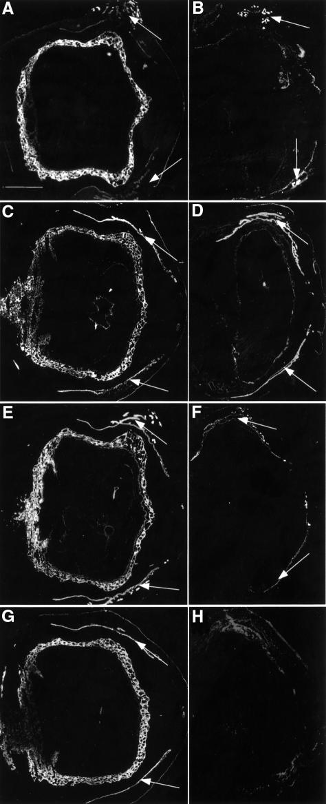
Fig. 2. Lack of K7, K18 and K19 in K18–/–K19–/– mice. (A and B) Immunofluorescence analysis of K7. (A) Strong staining of trophoblast giant cell layer in wild-type implantation sites. Maternal uterine glands and epithelia were stained weakly (arrows). (B) Positive staining of uterine epithelia and glands (arrows) but absence in doubly deficient extra-embyronic and embryonic compartments. (C and D) Staining for K8. (C) In wild-type animals, trophoblast giant cells, early placenta and maternal uterine glands and epithelia (arrows) were strongly positive. Parietal and visceral yolk sac, amnion, embryonic surface ectoderm, notochord and gut were also positive for K8. (D) Implantation site containing a doubly deficient embryo displayed strong staining in maternal uterine epithelia and glands (arrows). Weak staining was noted in the trophoblast giant cell layer and embryonic gut corresponding to K8 aggregates. (E and F) Staining for K18. (E) In wild type, the trophoblast giant cell layer and visceral yolk sac were strongly positive for K18. The amnion, embryonic ectoderm, gut and notochord were also positive. Maternal uterine glands and epithelia were stained. (F) Only maternal epithelia and glands were positive in doubly deficient implantation sites (arrows). (G and H) Staining for K19. (G) The trophoblast giant cell layer and maternal uterine epithelia were strongly positive. Weaker staining was observed in amnion, surface ectoderm, gut and notochord. (H) No staining was noted in doubly deficient embryos. The lack of staining in maternal tissues was due to the K19–/– genotype of parental mice. Bar, 1 mm.

An official website of the United States government
Here's how you know
Official websites use .gov
A
.gov website belongs to an official
government organization in the United States.
Secure .gov websites use HTTPS
A lock (
) or https:// means you've safely
connected to the .gov website. Share sensitive
information only on official, secure websites.
