
Figure 5.
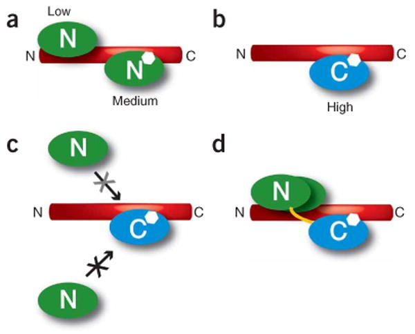
Schematic of Ca2+/CaM lobe multiple binding modes on the CaV1.2 IQ domain. (a) Ca2+/N lobe (green) has medium-affinity and low-affinity binding sites on the IQ domain. (b) Ca2+/C lobe (blue) has a high-affinity binding site on the IQ domain. (c) Binding of Ca2+/C lobe to the IQ domain blocks Ca2+/N lobe access to the Ca2+/N lobe medium-affinity site (black X) and reduces Ca2+/N lobe binding to the Ca2+/N lobe low-affinity site (grey X). (d) Representation of how Ca2+/C lobe binding to the IQ domain tethers Ca2+/N lobe near the Ca2+/N lobe low-affinity site. Yellow line indicates the CaM interlobe linker. In all panels, the approximate position of aromatic anchor Phe1628, which is shared by the Ca2+/N lobe medium-affinity site and the Ca2+/C lobe high-affinity site, is indicated by the white hexagon.