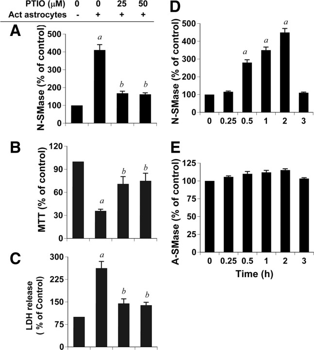
Figure 3.
Role of nitric oxide in activated astrocyte-induced cell death in neurons. Primary human astrocytes seeded in inserts were stimulated by the combination of Aβ and IL-1β as described above. After 24 h, media were removed and inserts were washed and placed on neurons that were pretreated with PTIO for 30 min. A, After 1 h, activity of N-SMase was measured in total cell extracts of neurons as described above. B, C, After 18 h, neuronal viability was examined by the metabolism of MTT (B) and the release of LDH (C). Values obtained from the control group served as 100%, and data obtained in other groups were calculated as a percentage of control accordingly. Results are the mean ± SD of three different experiments. ap < 0.001 versus control; bp < 0.001 versus activated astrocytes. D, E, Primary human neurons were treated with 25 μm DETA-NONOate (an NO donor), and at different time points of treatment, activities of N-SMase (D) and A-SMase (E) were monitored. ap < 0.001 versus control (0 h).

