FIGURE 1:
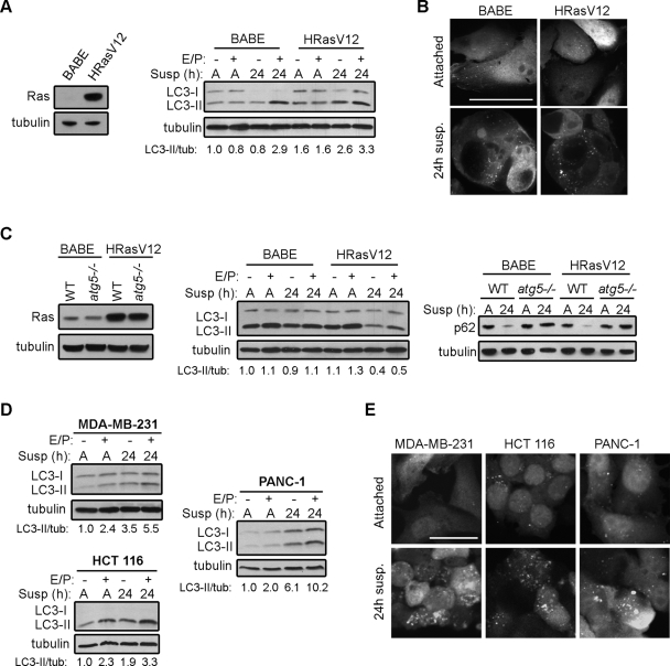
Oncogenic Ras does not suppress ECM detachment-induced autophagy. (A) Left: Ras expression in MCF10A cells expressing empty vector (BABE) or H-RasV12. Right: BABE and H-RasV12 MCF10A cells were grown attached (A) or suspended (susp) for the indicated times in the presence or absence of E64d and pepstatin A (E/P), lysed, and subjected to immunoblotting with antibodies against LC3 and tubulin. (B) GFP-LC3 puncta in MCF10A cells expressing empty vector (BABE) or H-RasV12 grown attached or suspended for 24 h. (C) Left: Ras expression in atg5+/+ (WT) and atg5−/− MEFs expressing empty vector or H-RasV12. Center: atg5+/+ (WT) MEFs expressing empty vector (BABE) and H-RasV12 were growth attached (A) or suspended (susp) for 24 h in the presence or absence of E64d and pepstatin A (E/P), lysed, and subjected to immunoblotting with antibodies against LC3 and tubulin. Right: atg5+/+ (WT) and atg5−/− MEFs expressing H-RasV12 or empty vector (BABE) were grown attached (A) or suspended (susp) for 24 h, lysed, and subjected to immunoblotting with antibodies against p62 and tubulin. (D) MDA-MB-231, HCT 116, and PANC-1 cells were grown attached (A) or suspended (susp) for 24 h in the presence or absence of E64d and pepstatin A (E/P) and subjected to immunoblotting with antibodies against LC3 and tubulin. (E) GFP-LC3 puncta in MDA-MB-231, HCT 116, and PANC-1 cells that were grown attached or detached for 24 h. Bar, 25 μm.

