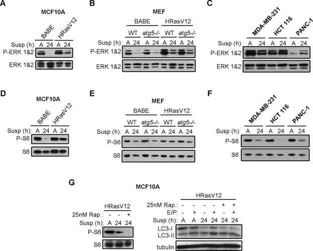
FIGURE 2:
Effects of ECM detachment on MAPK and mTORC1 signaling in Ras-transformed cells. (A–C) Empty vector (BABE) and H-RasV12–expressing MCF10A cells (A), atg5+/+ (WT) and atg5−/− MEFs (B), and K-Ras mutant carcinoma cell lines (C) were grown attached (A) or suspended (susp) for the indicated times and subjected to immunoblotting with antibodies against phosphorylated ERK1/2 and total ERK1/2 protein. (D–F) Empty vector (BABE) and H-RasV12–expressing MCF10A cells (D), atg5+/+ (WT) and atg5−/− MEFs (E), and K-Ras mutant carcinoma cell lines (F) were grown attached (A) or suspended (susp) for the indicated times and subjected to immunoblotting with antibodies against phosphorylated S6 and total ribosomal S6 protein. (G) H-RasV12 MCF10A cells were grown attached (A) or suspended (susp) for 24 h in the presence or absence of E64d and pepstatin A (E/P) and subjected to immunoblotting with antibodies against phosphorylated S6, S6, LC3, and tubulin. When indicated, cells were treated with 25 nM rapamycin for 5 h before harvest.

