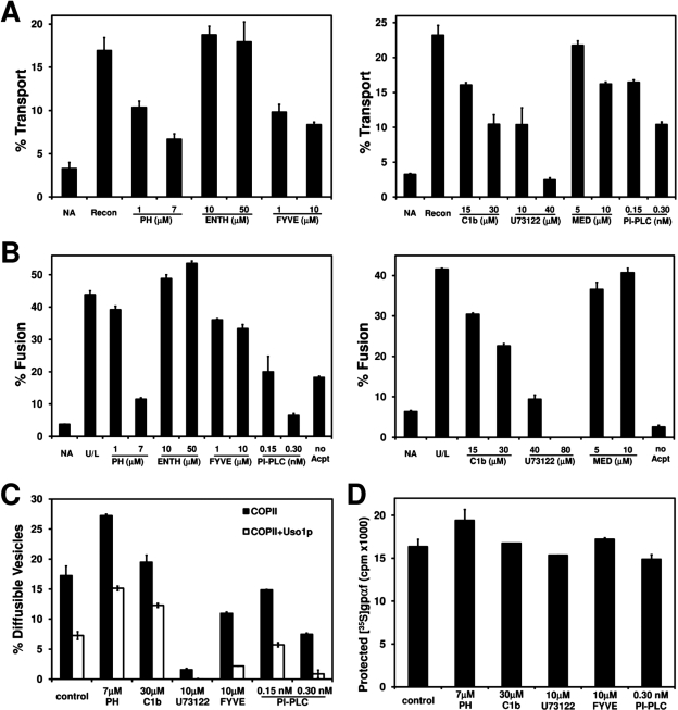
FIGURE 1:
Screen for inhibitors of ER-to-Golgi transport. (A) Washed semi-intact cells containing [35S]gpαf were pretreated with indicated inhibitors for 20 min at 4°C. Transport reactions were then incubated with Recon proteins (COPII, Uso1p, and LMA1) and an ATP regeneration system at 23°C for 1 h. The amount of Golgi-modified [35S]gpαf was measured to determine transport efficiency. NA is the background level of transport in the absence of transport factors. (B) Semi-intact cell acceptor membranes were pretreated with indicated inhibitors for 20 min at 4°C and then incubated with COPII vesicles containing [35S]gpαf in the presence of fusion factors (U/L: Uso1p and LMA1) and an ATP regeneration system at 23°C for 1 h. Golgi-modified [35S]gpαf was measured to determine fusion efficiency. The no Acpt condition represents the background level of fusion in the absence of semi-intact cell acceptor membranes. (C) Pretreated, semi-intact cells as in panel A were incubated with COPII or COPII plus Uso1p for 30 min at 23°C. To measure budding (black bars) or tethering (white bars), diffusible vesicles containing [35S]gpαf were separated from semi-intact cell membranes by centrifugation. (D) Mock-budding reactions as in panel C were treated with trypsin, and total protease protected [35S]gpαf was quantified to assess membrane integrity.

