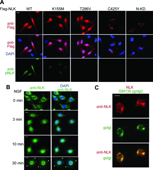
FIGURE 6:
Subcellular localization of exogenous and endogenous NLK proteins. (A) Dimerization of NLK is required for the nuclear localization. HeLa cells were transfected with Flag-NLK (WT), NLK(K155M), NLK(T286V), NLK(C425Y), and NLK(1–415) (N-KD) as indicated. Twenty-four hours after transfection, cells were fixed. Top panels show cells immunostained with anti-Flag antibody (red). Middle panels show cells stained with DAPI (blue) and anti-Flag antibody. Bottom panels show cells immunostained with anti-pNLK antibody (green). (B) NGF induces relocalization of NLK from the cytoplasm to the nucleus and leading edges. PC12 cells were treated with NGF for the indicated times and then fixed. PC12 cells were immunostained with anti-NLK antibody (green) and DAPI (blue). Arrowheads indicate the localization of NLK in the leading edge. Each panel shows one representative example from two repeated experiments. Scale bar (white line): 10 μm. (C) Endogenous NLK localizes to the Golgi in the absence of NGF stimulation. PC12 cells were fixed and immunostained with anti-NLK (red) and anti-GM130 (green) antibodies. Each panel shows one representative example from two repeated experiments.

