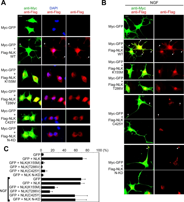
FIGURE 8:
Effects of NLK mutations on neurite induction. (A and B) PC12 cells were transfected with Myc-GFP, Flag-NLK (WT), NLK(K155M), NLK(T286V), NLK(C425Y), and NLK(1–415) (N-KD) as indicated. Twenty-four hours after transfection, cells were treated with (B) or without (A) 100 ng/ml NGF for 48 h. Cell shapes and nuclei were observed by staining with anti-Myc antibody (green) and DAPI (blue), respectively. Expression of the various NLK constructs, which were detected by anti-Flag antibody staining, is shown in red. NLK proteins in neurites are indicated with arrowheads. Each panel shows one or two representative examples from five repeated experiments. Scale bar (white line): 10 μm. (C) Quantification of neurite length. PC12 cells were transfected with expression plasmids as indicated. The lengths of the cellular processes or neurites were evaluated. Percentages of the cells with neurites/neurite-like processes are shown. The data shown represent the average of five independent experiments, and the error bars indicate the standard deviations.

