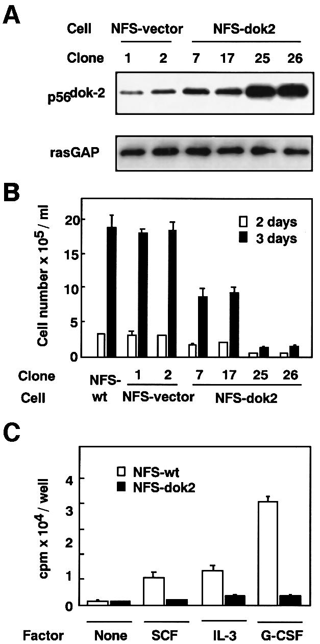
Fig. 2. Establishment of transfectants of M-NFS-60 cells expressing p56dok-2 and their proliferation rates. (A) M-NFS-60 cells were transfected with empty vector (NFS-vector) or with p56dok-2 cDNA (NFS-dok2). The transfectants were analyzed for their expression of p56dok-2 protein by immunoblotting. The M-NFS-60 cell lines were factor depleted for 4 h in RPMI 1640 medium containing 1% BSA and the cell lysates were prepared from the cells. The cleared cell lysates containing equal amounts of protein (5 µg/lane) were blotted to membrane and probed with anti-p56dok-2 antibody. To verify the amount of protein loaded, the same blot was probed with anti-rasGAP antibody. Data shown are representative of two independent experiments with similar results. (B) The proliferation of parental M-NFS-60 cells (NFS-wt), cells transfected with empty vector (NFS-vector) or cells transfected with p56dok-2 cDNA (NFS-dok2) in response to M-CSF. The cells were seeded at a density of 1 × 10⁴ cells/ml and cultured for 2 or 3 days in medium containing 10% FCS and M-CSF. Error bars from triplicate assays are shown. These results are representative of three independent experiments. (C) The proliferation of parental M-NFS-60 cells (open bars) or cells expressing p56dok-2 (clone 26, see A and B) (solid bars) cultured in the absence of factor (None), or the presence of SCF, IL-3 or G-CSF. Cells were seeded at a density of 2 × 10³ cells/well and cultured for 72 h. The cells were then pulsed with [3H]thymidine and the incorporated radioactivity was measured. Error bars from triplicate assays are shown. These results are representative of three independent experiments.
