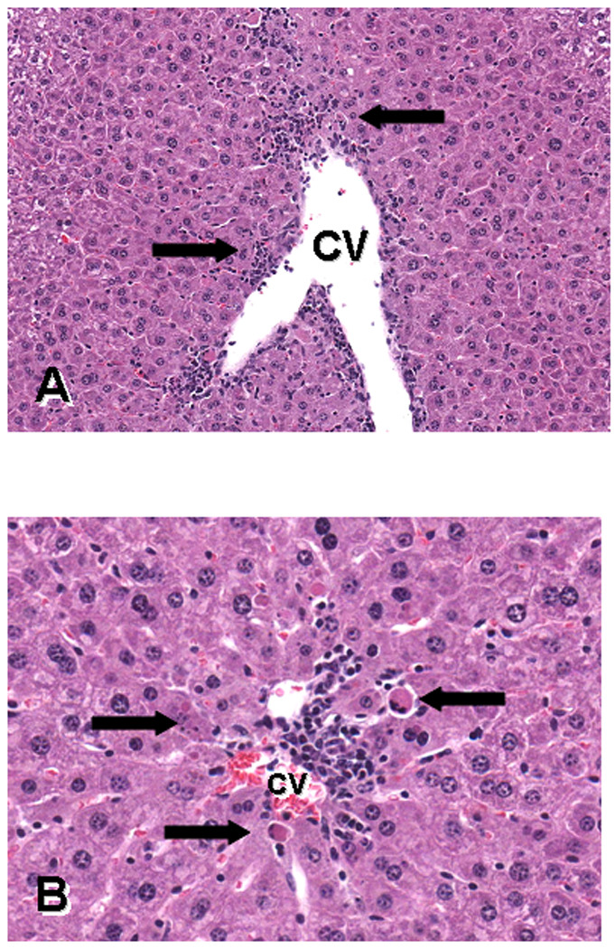
Fig. 2.

Photomicrographs of H&E sections of liver from a mouse treated with 50µg/kg of cylindrospermopsin during GD13–17 and killed on GD18, demonstrating centrilobular necrosis and apoptosis. The figures show different areas of the central vein (CV). Figure A magnification is 180X and B is 380X. The arrows in A point to cell death and inflammation; arrows in B point to apoptotic hepatocytes.
